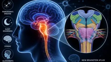
A team of Indian neuroscientists and computational engineers has achieved a major scientific milestone by releasing the world's most detailed 3D digital atlas of the human brainstem at cellular resolution. Developed at the Sudha Gopalakrishnan Brain Centre at IIT Madras, the "ANCHOR" platform maps over 200 brainstem nuclei, paving the way for advanced Alzheimer's and stroke clinical interventions.
CHENNAI, INDIA — In a major development for global neuromedicine, a multidisciplinary team of Indian neuroscientists, pathologists, and computational engineers has successfully mapped the human brain's least understood region at an unprecedented cellular resolution. The breakthrough establishes a new structural paradigm for diagnosing and treating degenerative neurological disorders.
Developed by the Sudha Gopalakrishnan Brain Centre (SGBC) at the Indian Institute of Technology Madras (IIT Madras), the newly released "ANCHOR" platform (Atlas of Neurochemical Characterization of the human brainstem with 3D Reconstruction) comprises the most comprehensive, multimodal 3D cellular maps of the human brainstem to date. Announcing the open-access data release contextually following the 3rd BRICS Neuroscience Symposium, official scientific bodies confirmed that the high-throughput multimodal whole-brain imaging pipeline allows researchers to inspect cellular topography that was previously invisible to modern medicine.
Mapping Over 200 Nuclei at Cellular Resolution
For more than a century, neuropathologists diagnosing complex cognitive disorders have inspected a handful of isolated tissue samples from an organ containing roughly 86 billion interconnected neurons. The ANCHOR platform bridges this historical visibility gap by creating a seamless structural transition from gross macro structures down to cellular level features.
According to data registries managed by the Press Information Bureau (PIB), the digital atlas covers the entire developmental lifespan from the prenatal stage through childhood and into adulthood. The structural framework maps out:
More than 200 complex brainstem nuclei and associated deep neural fiber tracts.
Hundreds of consecutive serial tissue sections processed through high-throughput imaging pipelines.
Eight separate, complementary neurochemical immunostains overlaid across 500 individual brain sections.
The brainstem acts as the primary physical bridge connecting the upper cerebrum to the spinal cord, controlling motor coordination, respiratory rhythms, and cardiovascular regulation. By utilizing advanced machine learning alignment algorithms, the SGBC has successfully matched cellular-level microscopy scans straight onto standard macroscopic Magnetic Resonance Imaging (MRI) coordinates.
Computational Convergence and Philanthropic Support
The development of the whole-brain imaging pipeline represents an intersection of biomedical science and advanced engineering talent in India. Operating with a global group of over 200 researchers and 20 international collaborators, the core engineering framework was initialized via state risk-capital grants and subsequently scaled up through private philanthropic backing from the Pratiksha Trust.
This institutional model runs parallel to a separate neuromorphic "moonshot" project simultaneously launched at the Indian Institute of Science (IISc). Also backed by the Pratiksha Trust, the IISc initiative is developing implantable and non-invasive "brain co-processors". These devices use artificial intelligence algorithms to decode neural recordings and re-encode signals via neurofeedback to rehabilitate stroke survivors, showcasing a broad national focus on indigenizing neurotechnologies.
Clinical Application in Degenerative Pathology
The immediate clinical objective of the ANCHOR atlas centers on isolating the cellular changes that occur during early-stage brain stem lesions. Medical teams at the center are already leveraging the 3D map database to study structural alterations induced by diseases such as Alzheimer's, dementia, and rabies.
By establishing a baseline map of a healthy human brainstem, researchers can now identify exactly which specific cell populations are targeted, distorted, or destroyed by progressive neuropathology. This cellular insight is critical for pharmaceutical laboratories designing targeted drug delivery molecules and clinicians seeking to identify cognitive diseases years before the appearance of visible physical symptoms.
Official Sources Section
The engineering specifications, data release parameters, and scientific project updates are sourced directly from research briefs authorized by the Sudha Gopalakrishnan Brain Centre (SGBC) and press updates managed by the Press Information Bureau (PIB). Cross-disciplinary data on brain co-processors conform to official engineering project logs released by the Indian Institute of Science (IISc).
Quote Section
Highlighting the strategic value of the digital asset release, Prof. V. Kamakoti, Director of IIT Madras, stated in a public announcement:
"This particular exploration puts IIT Madras in the frontiers of the most complex creation that this world has witnessed - the human brain. This Centre is also studying brains affected by different diseases like rabies, dementia and Alzheimer's disease. We now have a way by which we can say what happens to the basic structure of the brain due to diseases. This is a very important first step in understanding what happens in the human brain."
Why It Matters
For neurological patients and their families, the ANCHOR atlas accelerates the creation of precision diagnostics, turning diseases like Alzheimer's from late-stage surprises into highly manageable, early-tracked conditions. For AI developers and software engineers, the cellular-level wiring maps provide invaluable biological insights to design advanced, energy-efficient neuromorphic computing chips and artificial neural networks. Additionally, by offering this massive 3D database as a digital public good, India sets a global benchmark for open-science collaboration, significantly lowering the research costs associated with exploring the brain's remaining frontiers.
Key Facts at a Glance
Unprecedented Resolution: The ANCHOR platform maps more than 200 distinct brainstem nuclei at an explicit cellular level.
Multimodal Mapping: Successfully links fine, microscopic tissue sections with gross macroscopic clinical MRI coordinates.
Open Science Model: The comprehensive 3D data atlas has been made entirely available to global researchers in digital form.
Parallel Innovation: Complements an active moonshot project at IISc aimed at building AI-powered brain co-processors for stroke rehabilitation.
FAQ Section
What makes the ANCHOR atlas different from historical brain maps?
Traditional maps relied on isolated, two-dimensional tissue slides or low-resolution MRI scans. ANCHOR seamlessly integrates 3D computational reconstruction with multi-modal staining, allowing researchers to study cellular structures in full 3D context.
How will this brain mapping initiative aid in treating Alzheimer’s disease?
The digital atlas allows pathologists to compare diseased tissue against a normative, cellular-level baseline, identifying specific vulnerable cell populations and tracking exactly how structural patterns degrade over time.
Is the data generated by the Sudha Gopalakrishnan Brain Centre accessible to international scientists?
Yes. In line with public asset monetization and open-science policies, the 3D cell-resolution human brainstem atlas has been released publicly in a digital format for global scientific utility.
Source: Press Information Bureau (PIB); Indian Institute of Technology Madras (IIT Madras); Indian Institute of Science (IISc); Sudha Gopalakrishnan Brain Centre Research Registry.