


Loading market data...
ADVERTISEMENT

Latest Top News
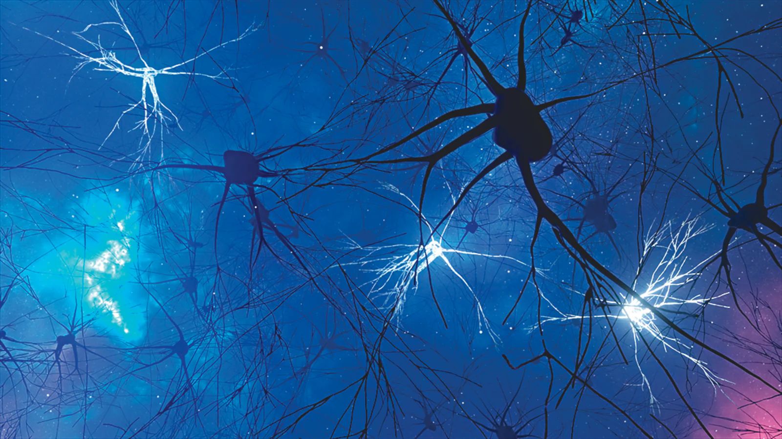
A Universe in a Cubic Millimeter: Scientists Peek Into the Brain’s Tiny Cosmos
In a groundbreaking decade-long project, neuroscientists from Harvard University, collaborating closely with Google Research experts, have mapped an unprecedentedly detailed 3D model of a tiny fragment—just 1 cubic millimeter—of human brain tissue. This microscopic piece, roughly half...
Stay Ahead – Explore Now! Three Generations Dimple Kapadia, Twinkle Khanna, Nitara Serve Fashion Goals at Airport
ADVERTISEMENT

Latest Updates

Spain vs Cabo Verde Match Result Ends in Historic...
16 Jun 2026, 06:01 AM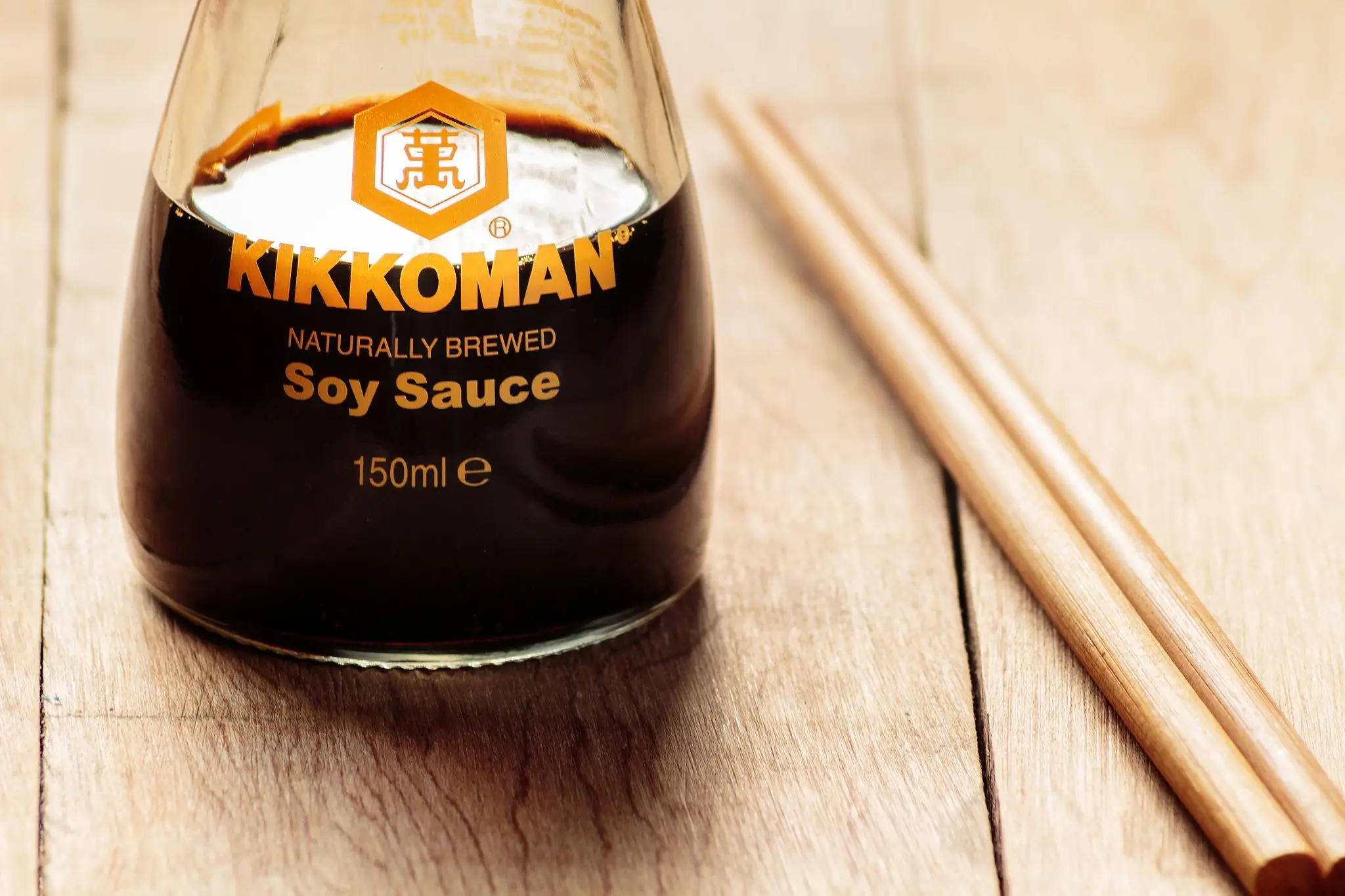
Kikkoman Plans First India Soy Sauce Manufacturing...
16 Jun 2026, 05:58 AM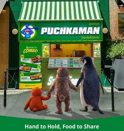
The Street Food Superhero: Puchkaman Scales 150% a...
16 Jun 2026, 12:11 AM
The Silence Value: Luxury Travellers Shift to Soft...
16 Jun 2026, 12:08 AM
The Great Tech Wake-Up Call: India Must Invest to...
16 Jun 2026, 12:01 AMADVERTISEMENT

Top Stories
Spain vs Cabo Verde Match Result Ends in Historic...
16 Jun 2026, 06:01 AM
Kikkoman Plans First India Soy Sauce Manufacturing...
16 Jun 2026, 05:58 AM
The Street Food Superhero: Puchkaman Scales 150% a...
16 Jun 2026, 12:11 AM