The Sudha Gopalakrishnan Brain Centre at IIT Madras has launched ANCHOR, the world's most detailed 3D digital atlas of the human brainstem at cellular resolution. Mapping over 200 structures across three lifespan stages, the open-source platform assists global clinicians in treating complex brain lesions and neurodegenerative disorders.
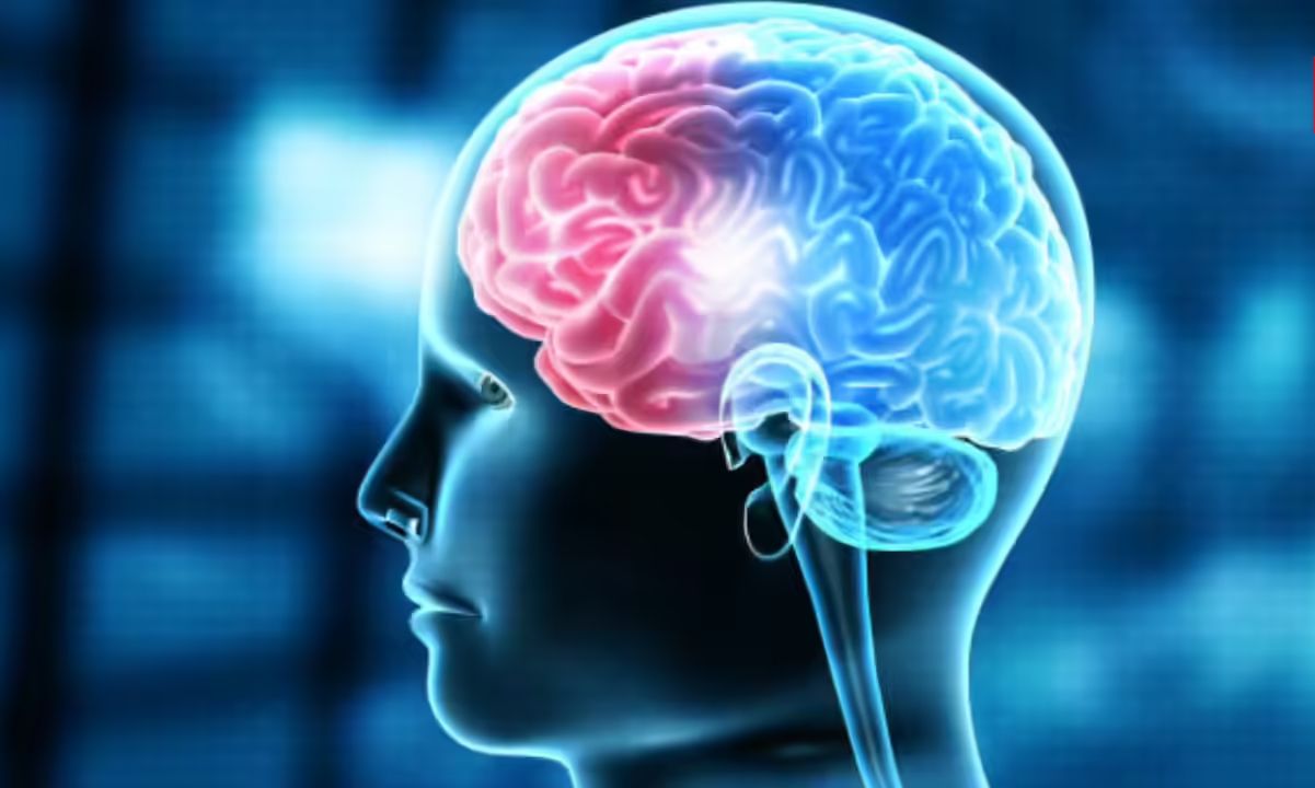
CHENNAI — The foundational biological structure governing human respiration, cardiac rhythm, and motor pathways has been mapped at single-cell resolution. The Indian Institute of Technology Madras (IIT Madras) has officially released the world’s most detailed three-dimensional digital atlas of the human brainstem, marking a significant leap forward for international neurobiology and clinical medicine.
Developed by the Sudha Gopalakrishnan Brain Centre (SGBC) at IIT Madras, the open-source platform—titled ANCHOR (Atlas of Neurochemical Characterization of the Human Brainstem with 3D Reconstruction)—presents the most complete multi-modal architectural map of this complex nerve center to date. The structural database spans key developmental stages, stretching from the prenatal period to childhood and full adulthood.
Unveiled on campus at the 3rd BRICS Neuroscience Symposium, technology administrators emphasized that the launch represents a crucial turning point for modern medicine. By integrating macro-scale volumetric MRI data directly with micro-scale cellular images, the open-access atlas gives global researchers a reliable map to locate specific cell populations damaged by traumatic brainstem injuries and long-term neurodegenerative diseases.
The Neurochemical Architecture of the ANCHOR Initiative
The mapping project used advanced high-throughput histology pipelines to convert post-mortem physical brain tissue into an exploreable petabyte-scale 3D volume.
According to technical specifications published on the official ANCHOR Web Portal, the system resolves tiny cell categories that have never been captured clearly in past digital atlases. By overlaying eight distinct neurochemical stains across a sequence of more than 500 microscopic slices, the engineering and medical research teams successfully isolated unique cell groups across vital centers, including the midbrain, pons, and medulla oblongata. This extreme precision bridges a massive data gap in neuroanatomy, where traditional clinical scans remain too blurry to display individual cell changes.
Bridging Engineering Talents and Clinical Neuroscience
The cross-disciplinary development represents a landmark victory for deep-tech medical research frameworks inside India's higher education systems.
Seamless Dual-Scale Imaging Navigation
Medical experts point out that the main technical challenge in brain mapping has always been maintaining accurate alignment when zooming from whole-organ scans down to microscopic cell fields.
According to institutional design briefs released by IIT Madras, the project succeeded by combining three distinct structural datasets into one interactive workspace:
Macro-Scale Realignment: Utilizing high-resolution structural post-mortem MRI to define the core outer volumetric shape of the specimen.
Micro-Scale Cellular Overlay: Digitizing high-magnification tissue sections to identify catecholaminergic cell groups and intricate dendritic patterns.
Lifespan Comparative Timelines: Stacking complete baseline files for a 25-week gestational fetus, a 9-year-old child, and a 54-year-old adult.
Cross-Institutional Sourcing: Collaborating with domestic medical bodies—such as CMC Vellore and Kilpauk Medical College—to safely and ethically acquire tissue samples.
Impact on Neurodegenerative Disease Research
Independent scientific reviewers note that mapping the exact boundaries of the brainstem helps clarify how aggressive viral or age-related diseases degrade essential human functions.
"According to research statements distributed by the Principal Scientific Adviser to the Government of India, this multimodal architecture marks an exceptional milestone in neurobiology. The open digital availability of these maps will assist clinical teams globally in identifying the exact cell subgroups destroyed during brainstem lesions, helping doctors predict patient recovery timelines much more accurately."
The research center has already moved into its next phase, studying how severe conditions like dementia, rabies, and Alzheimer’s disease structurally damage these freshly mapped zones. This long-term dataset will help international pharmaceutical firms test whether targeted drug therapies are successfully protecting vulnerable cells at the microscopic level.
Why It Matters
For practicing neurosurgeons and clinical oncologists, this cell-level resolution atlas removes a great deal of guesswork when navigating delicate surgeries around brainstem lesions. Having an exact structural roadmap helps surgical teams preserve healthy fiber tracts, lowering the risk of paralyzing motor nerves during deep-tissue procedures.
For global neuroscience teams, the open-source release of this structural data completely changes how labs study human development. By offering a highly detailed baseline of healthy tissue across a lifespan, the platform allows international researchers to study complex sleep, respiratory, and balance disorders without needing to build expensive imaging pipelines of their own.
Key Facts at a Glance
Global Mapping Record: IIT Madras has built the world's most detailed 3D cell-resolution atlas of the human brainstem, mapping over 200 structures.
The Technology Platform: The ANCHOR model integrates gross volumetric MRI scans with cellular-level tissue slides to allow seamless zooming.
Lifespan Documentation: The open-access resource tracks structural changes from the second trimester of pregnancy up into childhood and mature adulthood.
The Collaborative Network: The project brought together a global team of over 200 engineers, technicians, and neuroscientists working alongside 20 international partners.
Frequently Asked Questions
How can global researchers access the new 3D human brainstem dataset?
The entire multi-modal map is open to the public and can be explored interactively through the official web domain at https://anchor.humanbrain.in/.
Why did the research team prioritize mapping the brainstem over other brain regions?
The brainstem serves as the primary bridge between the brain and the spinal cord. It controls all motor movements and vital life functions like breathing, sleeping patterns, and blood pressure.
What makes the ANCHOR atlas more detailed than previous medical models?
Unlike standard MRI scans that only show overall brain shapes, this project combines deep tissue staining with microscopic imaging, allowing researchers to study individual cells.
Official Sources Section: